
Gene Expression of Subtilisin Genes of UV-irradiated Trichophyton indotineae Isolated from Iraqi Patients
DOI:
https://doi.org/10.32007/jfacmedbaghdad3116Keywords:
Dermatophytes, Gene expression, Subtilisin genes, Trichophyton indotineae, UV radiationAbstract
Background: Virulence genes in dermatophytes have been a major focus of research in recent years. These fungi have a unique ability to degrade keratin, an essential structural protein in human skin, using protease enzymes. These enzymes play a crucial role in the development and spread of infections.
Objectives: To assess the impact of UV radiation on the expression of subtilisin virulence genes in Trichophyton indotineae isolates obtained from patients in Baghdad, Iraq.
Materials: Seven T. indotineae isolates were analyzed in this study, and their proteolytic activity was qualitatively assessed using skim milk agar. The isolate displaying the largest clearance zone in response to UV radiation (254–365 nm) was selected for further experiments with varying exposure durations. The expression of Subtilisin (SUB) genes was measured using the one-step RT-qPCR technique. These specimens were obtained from patients diagnosed with Tinea at Al-Yarmouk Teaching Hospital and Al-Zahra Consultative Center for Allergy and Asthma in Baghdad, and the laboratory procedures were conducted in the Department of Biology, College of Science, University of Baghdad, between October 2022 and February 2023.
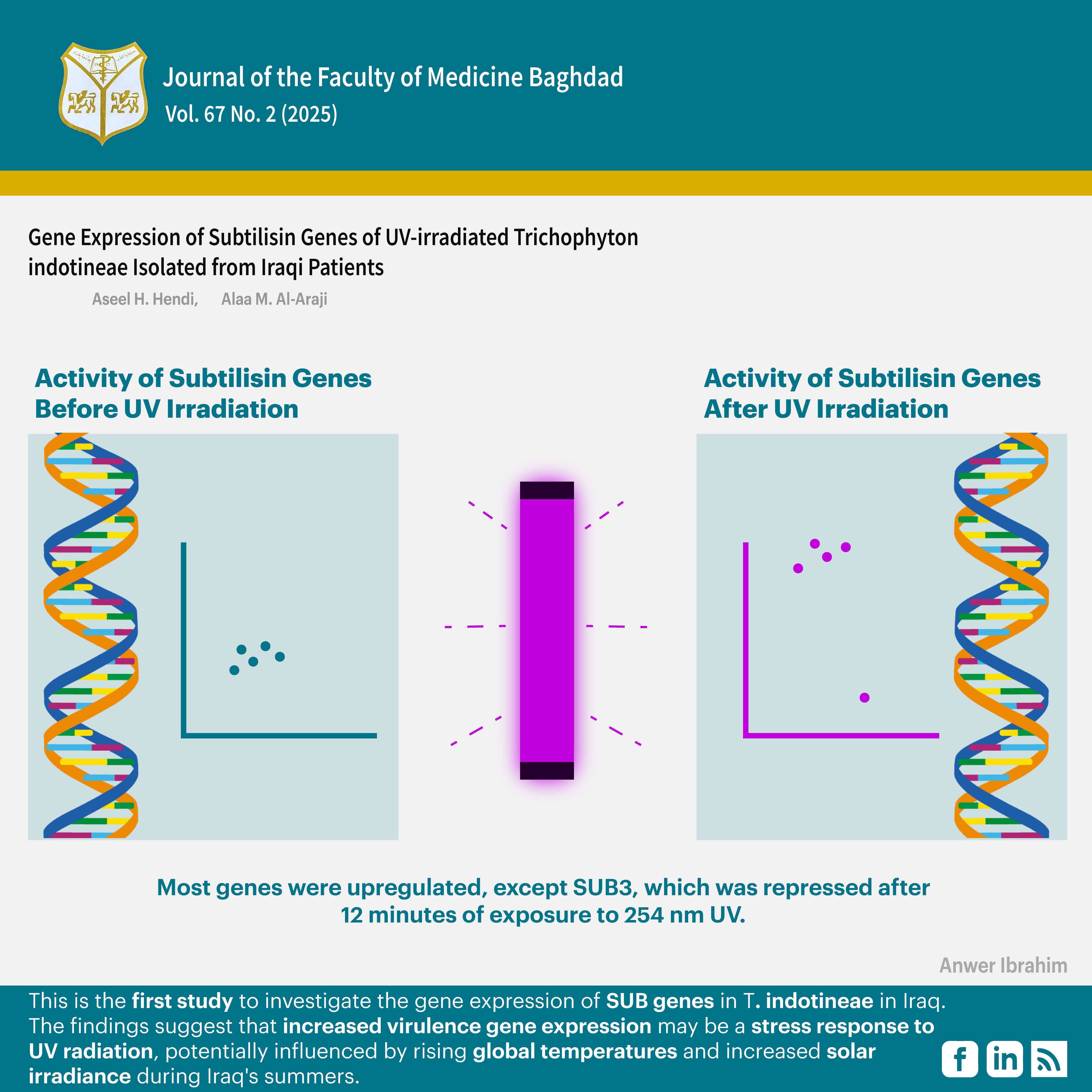
Results: Distinct trends in SUB7 gene expression were observed following UV exposure. A significant increase was noted after 6 minutes of exposure to 365 nm UV, while a rapid rise was seen after 3 minutes of exposure to 254 nm UV. All the studied genes (SUB1, SUB3, SUB4, SUB6, and SUB7) exhibited higher expression levels following UV exposure for 3 or 6 minutes. Most genes were upregulated, except SUB3, which was repressed after 12 minutes of exposure to 254 nm UV.
Conclusion: The findings suggest that increased virulence gene expression may be a stress response to UV radiation, potentially influenced by rising global temperatures and increased solar irradiance during Iraqi summers.
Received: Jan. 2025
Revised: June 2025
Accepted: June 2025
Published Online: June 2025
Published: July 2025
References
1. Cañete-Gibas CF, Mele J, Patterson HP, Sanders CJ, Ferrer D, Garcia V, et al. Terbinafine-Resistant Dermatophytes and the Presence of Trichophyton indotineae in North America. Journal of Clinical Microbiology.2023;61(8):e0056223.https://doi.org/10.1128/jcm.00562-23
2. Ebert A, Monod M, Salamin K, Burmester A, Uhrlaß S, Wiegand C, et al. Alarming India‐wide phenomenon of antifungal resistance in dermatophytes: A multicentre study. Mycoses. 2020;17;63(7):717-28.https://doi.org/10.1111/myc.13091
3. Khan S M, Farhana A, Amin U, Khan RA. Clinicomycological pattern of dermatomycosis – A two-year retrospective study in a tertiary care hospital of north India. Indian J Microbiol Res 2021;8(1):58-64.https://doi.org/10.18231/j.ijmr.2021.013
4. Ramos MLM, Coelho RA, Brito-Santos F, Guimarães D, Premazzi M, Zancopé-Oliveira RM, et al. Comparative Analysis of Putative Virulence-Associated Factors of Microsporum canis Isolates from Human and Animal Patients. Mycopathologia.2020;185(4):665–73. https://doi.org/10.1007/s11046-020-00470-9
5. Hernández BC, Rives IM, Manrique BA, Aragües IH, Gómez CL, Cazaña TG. Comment on: Report of terbinafine‐resistant Trichophyton indotineae in a pregnant patient—A diagnostic and therapeutic challenge. Journal of the European Academy of Dermatology and Venereology. 2023;38(7).https://doi.org/10.1111/jdv.19672
6. Herrera EM, Coutiño GM, Venado CEF, Castro HR, Arenas R, Almazán RP, et al. Main Phenotypic Virulence Factors Identified in Trichophyton rubrum. Journal of Biological Regulators and Homeostatic Agents. 2023; 37(5): 2345-2356 https://doi.org/10.23812/j.biol.regul.homeost.agents.20233705.231
7. Bitencourt TA, Lang EAS, Sanches PR, Peres NTA, Oliveira VM, Fachin AL, et al. HacA Governs Virulence Traits and Adaptive Stress Responses in Trichophyton rubrum. Frontiers in Microbiology. 2020;11. https://doi.org/10.3389/fmicb.2020.00193
8. Ferrall-Fairbanks MC, Kieslich CA, Platt MO. Reassessing enzyme kinetics: Considering protease-as-substrate interactions in proteolytic networks. Proceedings of the National Academy of Sciences. 2020;117(6):3307–18. https://doi.org/10.1073/pnas.1912207117
9. Baumbach C, Michler JK, Nenoff P, Uhrlaß S, Schrödl W. Visualising virulence factors: Trichophyton benhamiaes subtilisins demonstrated in a guinea pig skin ex vivo model. Mycoses. 2020;63(9):970–8. https://doi.org/10.1111/myc.13136
10. Dellière S, Jabet A, Abdolrasouli A. Current and emerging issues in dermatophyte infections. PLoS Pathogens. 2024;20(6):e1012258. https://pmc.ncbi.nlm.nih.gov/articles/PMC11175395/
11. Baumbach CM, Rückner A, Partusch L, Engel E, Schrödl W, Michler JK. Comprehensive Assessment of the Virulence Factors sub 3, sub 6 and mcpA in the Zoonotic Dermatophyte Trichophyton benhamiae Using FISH and qPCR. Journal of Fungi. 2021;8(1):24.https://doi.org/10.3390/jof8010024.
12. Martins-Santana L, Petrucelli MF, Sanches PR, Martinez-Rossi NM, Rossi A. Peptidase Regulation in Trichophyton rubrum Is Mediated by the Synergism Between Alternative Splicing and StuA-Dependent Transcriptional Mechanisms. Frontiers in Microbiology. 2022;13. https://doi.org/10.3389/fmicb.2022.930398
13. Chen J, Gao Y, Xiong S, Peng Z, Zhan P. Expression Profiles of Protease in Onychomycosis-Related Pathogenic Trichophyton rubrum and Tinea Capitis-Related Pathogenic Trichophyton violaceum. Mycopathologia. 2024;189(1).
https://doi.org/10.1007/s11046-024-00828-3
14. Baumbach CM, Rückner A, Partusch L, Engel E, Schrödl W, Michler JK. Comprehensive Assessment of the Virulence Factors sub 3, sub 6 and mcpA in the Zoonotic Dermatophyte Trichophyton benhamiae Using FISH and qPCR. Journal of Fungi. 2021;8(1):24.https://doi.org/10.3390/jof8010024
15. Bohacz J, Możejko M. Keratinolytic activity of pigmenting and non-pigmenting soils strains of Trichophyton ajelloi. International Biodeterioration & Biodegradation. 2023;186:105704.
https://doi.org/10.1016/j.ibiod.2023.105704
16. Gautham RC, Suhana SK, Kumar JN, Devi VK, Ulitappa G, Hasini B, et al. A cross-sectional clinicomycological study of tinea corporis in an indian tertiary care hospital. International Journal of Clinical Pharmacokinetics and Medical Sciences. 2024;4(3):28–37. https://doi.org/10.26452/ijcpms.v4i3.650
17. Al-Araji AM, Al-Jboury MH, Aboud HM. Induce mutations for Bavistin resistance in Trichoderma harzianum by UV-irradation. Iraqi Journal of Science. 2013;54(5). http://www.ijs.scbaghdad.edu.iq/issues/Vol54/No5/Vol54Y2013No5P1023-1035.pdf
18. Nematollahi AR, Badiee P, Nournia E. The efficacy of ultraviolet irradiation on trichophyton species isolated from nails. Jundishapur Journal of Microbiology. 2015;8(6). https://doi.org/10.5812/jjm.18158v2
19. Rio DC, Ares M, Hannon GJ, Nilsen TW. Purification of RNA using TRIZol (TRI reagent). Cold Spring Harbor Protocols. 2010;6:5439. https://doi.org/10.1101/pdb.prot5439
20. Khedmati E, Hazaveh SJH, Bayat M, Amini K. Identification of subtilisin virulence genes (SUB1-7) in Epidermophyton floccosum isolated from patients with dermatophytosis in Iran. Gene Reports. 2020;20:100748. https://doi.org/10.1016/j.genrep.2020.100748
21. Jacob TR, Peres NTA, Persinoti GF, Silva LG, Mazucato M, Rossi A, et al. rpb2is a reliable reference gene for quantitative gene expression analysis in the dermatophyte Trichophyton rubrum. Medical Mycology. 2012 ;50(4):368–77. https://doi.org/10.3109/13693786.2011.616230
22. Hashoosh QH, Al-Aaraji AMohsin. Metalloprotease Genes Expression in Trichophyton mentagraphytes and Trichophyton simii Contains Genetic Variations Isolated from Iraqi Patients Resistance to Ketoconazole and Amphotericin B. IOP Conference Series Earth and Environmental Science.2024;1325(1):012025. https://doi.org/10.1088/1755-1315/1325/1/012025
23. Dubljanin E, Zunic J, Vujcic I, Calovski IC, Grujicic SS, Mijatovic S, et al. Host-Pathogen interaction and resistance mechanisms in dermatophytes. Pathogens. 2024;13(8):657. https://doi.org/10.3390/pathogens13080657.
24. Mahmood HR, Shams-Ghahfarokhi M, Salehi Z, Razzaghi-Abyaneh M. Epidemiological trends, antifungal drug susceptibility and SQLE point mutations in etiologic species of human dermatophytosis in Al-Diwaneyah, Iraq. Scientific Reports. 2024;14(1). https://doi.org/10.1038/s41598-024-63425-w
25. Kubaisi T. Resistant dermatophytosis: a stubborn habit and major challenge in Iraq. Al- Anbar Medical Journal. 2023; 19(2): 78–80..https://doi.org/10.33091/amj.2023.141295.1225
26. Wong HJ, Mohamad-Fauzi N, Rizman-Idid M, Convey P, Alias SA. Protective mechanisms and responses of micro-fungi towards ultraviolet-induced cellular damage. Polar Science. 2018;20:19–34. https://doi.org/10.1016/j.polar.2018.10.001
27. Çavuşoğlu K, Macar TK, Macar O, Çavuşoğlu D, Yalçın E. Comparative investigation of toxicity induced by UV-A and UV-C radiation using Allium test. Environmental Science and Pollution Research. 2022;29(23):33988–98. https://doi.org/10.1007/s11356-021-18147-1.
28. Gao P, Jin K, Xia Y. The phosphatase gene MaCdc14 negatively regulates UV-B tolerance by mediating the transcription of melanin synthesis-related genes and contributes to conidiation in Metarhizium acridum. Current Genetics. 2019;66(1):141–53.
https://doi.org/10.1007/s00294-019-01008-
30.Mohammad TH, Habeb KA. Epidemiological study of keratinophilic fungi in Baghdad swimming pools. Baghdad Science Journal. 2014;11(3):1122–9. https://doi.org/10.21123/bsj.2014.11.3.1122-1129
Downloads
Published
Issue
Section
Categories
License
Copyright (c) 2025 Aseel H. Hendi, Alaa Al-Araji

This work is licensed under a Creative Commons Attribution 4.0 International License.


 Creative Commons Attribution 4.0 International license..
