Assessment of Serum P53 Protein Level in Adult Patients with Acute Myeloid Leukemia in Correlation with Response to Treatment
DOI:
https://doi.org/10.32007/jfacmedbaghdad.6632345الكلمات المفتاحية:
AML، ELISA، ; Remission، Survival status، 53.الملخص
Background: Acute myeloid leukemia (AML) is an adult leukemia characterized by rapid proliferation of undifferentiated myeloid precursors, leading to bone marrow (BM) failure and impaired erythropoiesis. The p53 tumor suppressor protein regulates cell division and inhibits tumor development by preventing cell proliferation of altered or damaged DNA. It orchestrates various cellular reactions, including cell cycle arrest, DNA repair, and antioxidant properties.
Objectives: To investigate the relationship of P53 serum level with hematological findings, remission, and survival status in de novo AML patients.
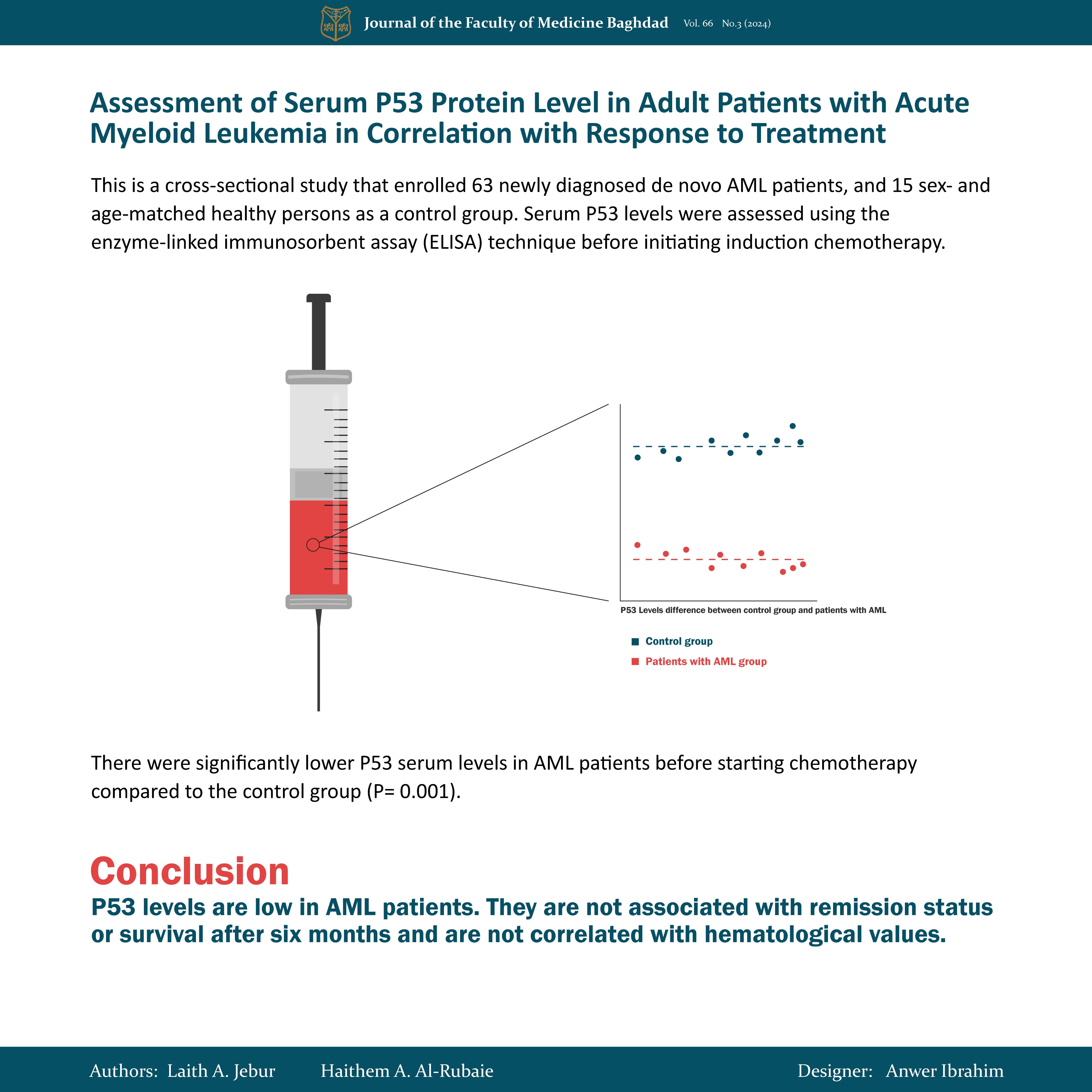
Methods: This is a cross-sectional study that enrolled 63 newly diagnosed de novo AML patients, and 15 sex- and age-matched healthy persons as a control group. Serum P53 levels were assessed using the enzyme-linked immunosorbent assay (ELISA) technique before initiating induction chemotherapy. The study was performed between November 2022 and May 2023 at the Hematology and Bone Marrow Transplant Center of the Medical City Complex in Baghdad.
Results: There were significantly lower P53 serum levels in AML patients before starting chemotherapy compared to the control group. However, no substantial difference in P53 levels was identified between AML patients achieving complete remission and those exhibiting no response, nor between alive and deceased individuals. Furthermore, there was a positive yet statistically non-significant correlation between serum P53 levels and age, and no significant relationship between P53 levels and sex or various hematological parameters.
Conclusion: P53 levels are low in AML patients. They are not associated with remission status or survival after six months and are not correlated with hematological values.
التنزيلات
المراجع
Dawood HH, Mohammed RK. The Correlation Study between TP53 Gene Expression and Acute Myeloid Leukemia in Iraq. Iraqi Journal of Science. 2023:5615-23.https://doi.org/10.24996/ijs.2023.64.11.14.
Borrero LJH, El-Deiry WS. Tumor suppressor p53: Biology, signaling pathways, and therapeutic targeting. Biochimica et Biophysica Acta (BBA)-Reviews on Cancer. 2021;1876(1):188556. https://doi.org/10.1016/j.bbcan.2021.188556.
Boutelle AM, Attardi LD. p53 and tumor suppression: it takes a network. Trends in cell biology. 2021;31(4):298-310. https://doi.org/10.1016/j.tcb.2020.12.011.
George B, Kantarjian H, Baran N, Krocker JD, Rios A. TP53 in acute myeloid leukemia: molecular aspects and patterns of mutation. International journal of molecular sciences. 2021;22(19):10782. https://doi.org/10.3390/ijms221910782.
Heuser M, Ofran Y, Boissel N, Mauri SB, Craddock C, Janssen J, et al. Acute myeloid leukaemia in adult patients: ESMO Clinical Practice Guidelines for diagnosis, treatment and follow-up. Annals of Oncology. 2020;31(6):697-712https://doi.org/10.1016/j.annonc.2020.02.018.
AL-Saidi DN, Hameed BM, Khaleel KJ. Detection of RUNX1-RUNX1T1 Fusion Gene in AML Patients by FISH Technique in Iraq. Journal of Communicable Diseases (E-ISSN: 2581-351X & P-ISSN: 0019-5138). 2021;53(4):54-60. https://doi.org/10.24321/0019.5138.202174.
Mahmood EF, Ahmed AA. Evaluation of interleukin-35 and interleukin-10 in adult acute myeloid leukemia patients before and after induction chemotherapy. Iraqi Journal of Hematology. 2020;9(2):82-6. https://doi.org/10.4103/ijh.ijh_17_20.
Hamad HM, Shabeeb ZA, Awad MM. Expressions of CD274 (PD-L1) and CD47 receptors on the surface of blast cells in AML patients. Iraqi Journal of Science. 2022:2373-87. https://doi.org/10.24996/ijs.2022.63.6.6.
Kayser S, Hills RK, Langova R, Kramer M, Guijarro F, Sustkova Z, et al. Characteristics and outcome of patients with acute myeloid leukaemia and t (8; 16)(p11; p13): results from an International Collaborative Study. British journal of haematology. 2021;192(5):832-42. https://doi.org/10.1111/bjh.17336.
Koolivand M, Ansari M, Moein S, Afsa M, Malekzadeh K. The inhibitory effect of sulforaphane on the proliferation of acute myeloid leukemia cell lines through controlling miR-181a. Cell Journal (Yakhteh). 2022;24(1):44. https://doi.org/10.22074/CELLJ.2022.7508.
El-Toukhy MM, Morad HAM, Hossam AH, Farrag WFM. Serum Epidermal Growth Factor Receptor and p53 in Patients with Acute Myeloid Leukemia. The Medical Journal of Cairo University. 2019;87(June):1363-9. https://doi.org/10.21608/mjcu.2019.53428.
Mallouh SS, Alawadi NB, Hasson AF. Serum vascular endothelial growth factor levels in Iraqi patients with newly diagnosed acute leukemia. Medical Journal of Babylon. 2015;12(1):21.
https://www.uobabylon.edu.iq/publications/medicine_edition24/medicine24_26.doc.
Moulod SH, AL-Rubaie HA. BCMA plasma level: its relation to induction therapy response in adult acute myeloid leukemia. Biochem Cell Arch. 2022;22(1):895-9. Doc ID: https://connectjournals.com/03896.2022.22.895.
Al-Bayaa IM, Al-Rubaie HA, Al-Shammari HH. Evaluation of Hepatocyte Growth Factor in Iraqi Patients with Acute Myeloid Leukemia: Its Correlation with Clinical Parameters and Response to Induction Therapy: its correlation with clinical parameters and response to induction therapy. Open Access Macedonian Journal of Medical Sciences (OAMJMS). 2020;8(B):49-53. https://doi.org/10.3889/oamjms.2020.4235.
Abd MS, Alwash MM, Ahmed AA. Polymorphism of TET2 gene among Iraqi acute myeloid leukemia. Biochem Cell Arch. 2020;20(2):4571-5. https://doi.org/10.13140/RG.2.2.21268.32642.
Venkatesan S, Boj S, Nagaraj S. A Study of clinico-hematological profile in acute leukemia with cytochemical correlation. Int J Acad Med Pharm. 2023;5(4):893-8. https://doi.org/10.47009/jamp.2023.5.4.181.
Hasham S, Taj AS, Masood T, Haq M. Frequency of FAB subtype and clinicohaematological manifestation in elderly acute myeloid leukemia patients in tertiary care hospitals Peshawar. Sch J App Med Sci. 2022;9:1425-30. https://doi.org/10.36347/sjams.2022.v10i09.002.
Alwan AF, Zedan ZJ, Salman OS. Acute myeloid leukemia: clinical features and follow-up of 115 Iraqi patients admitted to Baghdad Teaching Hospital. Tikrit Med J. 2009;15(1):1-8.
https://scholar.google.com/scholar?cluster=9120028953813627648&hl=ar&as_sdt=0,5.
Zayed RA, Eltaweel MA, Botros SK, Zaki MA. MN1 and PTEN gene expression in acute myeloid leukemia. Cancer Biomarkers. 2017;18(2):177-82. https://doi.org/10.3233/CBM-160235.
Moulod SH, AL-Rubaie HA. APRIL mRNA expression: does it predict the response to induction therapy in adult acute myeloid leukemia? Biochem Cell Arch. 2022;22(1):957-61. DocID: https://connectjournals.com/03896.2022.22.957.
Udupa MN, Babu KG, Babu MS, Lakshmaiah K, Lokanatha D, Jacob AL, et al. Clinical profile, cytogenetics and treatment outcomes of adult acute myeloid leukemia. Journal of cancer research and therapeutics. 2020;16(1):18-22. https://doi.org/10.4103/jcrt.JCRT_1162_16.
Sadek NA, Abd-eltawab SM, Assem NM, Hamdy HA, EL-sayed FM, Ahmad MA-R, et al. Prognostic value of absolute lymphocyte count, lymphocyte percentage, serum albumin, aberrant expression of CD7, CD19 and the tumor suppressors (PTEN and p53) in patients with acute myeloid leukemia. Asian Pacific Journal of Cancer Biology. 2020;5(4):131-40.
https://doi.org/10.31557/apjcb.2020.5.4.131-140.
Abdel-Aziz MM. Clinical significance of serum p53 and epidermal growth factor receptor in patients with acute leukemia. Asian Pacific Journal of Cancer Prevention. 2013;14(7):4295-9. https://doi.org/10.7314/APJCP.2013.14.7.4295.
McKeon FD. p63 and p73 in tumor suppression and promotion. Cancer Research and Treatment: Official Journal of Korean Cancer Association. 2004;36(1):6. https://doi.org/10.4143/crt.2004.36.1.6.
التنزيلات
منشور
إصدار
القسم
الرخصة
الحقوق الفكرية (c) 2024 Laith A. Jebur, Haithem A. Al-Rubaie

هذا العمل مرخص بموجب Creative Commons Attribution 4.0 International License.









 Creative Commons Attribution 4.0 International license..


